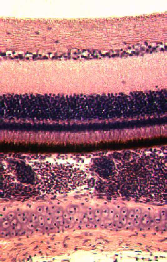
Left photograph: completely unpigmented area in the choroid of the eye of a recessive pied (s / s) Budgerigar
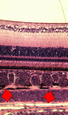
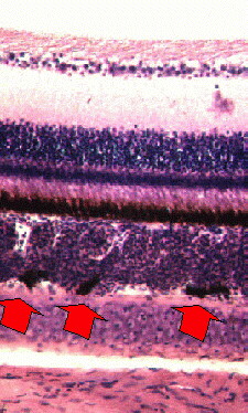
Middle and right photographs: poorly pigmented areas in the choroid of the eye of a recessive pied (s / s) Budgerigar
Note red arrows pointing at some abnormal melanocytes in between the sclera and the choroid.